Auricular microsystem
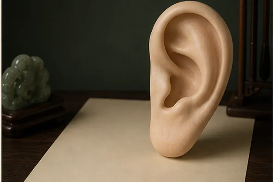
A comparative introduction to Chinese auricular nomenclature, European cartographies, and visible anatomical references.
History
Ear references appear in early Chinese texts; modern somatotopic mapping developed and was standardized in the twentieth century. Schools are not identical.
The map model
The “inverted fetus” model is a mnemonic, but points should be studied by anatomy, nomenclature, and the specific standard used.
Regions of the map
Curved outer rim of the auricle.
Rim points and surface references.
Learn the root, tubercle, and tail of the helix before memorizing points.
Inner ridge dividing into two crura.
Maps of trunk and limbs.
Distinguish body, superior crus, and inferior crus.
Central depression around the ear canal.
Cavity and internal-system references in several standards.
Separate cymba and cavum using the helix root.
Anterior and inferior prominences facing the concha.
Head, regulatory, and sensory references depending on school.
Identify supratragic and intertragic notches.
Soft inferior portion without cartilage.
Maps of face, mouth, and sensory organs.
Use a grid only as a convention of the standard being studied.
What the evidence says
Clinical research varies by indication, protocol, and methodological quality. An ear map does not diagnose from color, tenderness, or texture changes.
Safety
Assessment and invasive stimulation belong to trained professionals. Do not insert needles, seeds, or devices over damaged skin, and do not use the ear for self-diagnosis.
Health notice. Educational and study content. It describes concepts of Traditional Chinese Medicine and does not replace diagnosis or treatment by a healthcare professional. Needling, medicinal herbs and invasive techniques must be applied by trained professionals. Safety guide.